Subscribe to get full access to this operation and the extensive Upper Limb & Hand Surgery Atlas.
FREE TRIAL
Professional Guidelines Included
Learn the Distal radial fracture: Dorsal plating with Depuy/Synthes 24mm VA locking radial column plate assisted by wrist arthroscopy using Acumed ARC tower surgical technique with step by step instructions on OrthOracle. Our e-learning platform contains high resolution images and a certified CME of the Distal radial fracture: Dorsal plating with Depuy/Synthes 24mm VA locking radial column plate assisted by wrist arthroscopy using Acumed ARC tower surgical procedure.
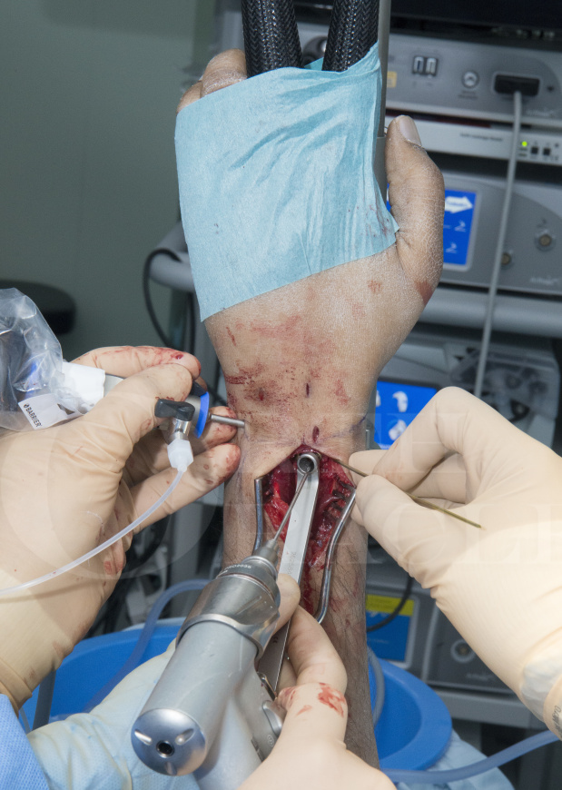
This is a detailed step by step instruction through a arthroscopic assisted dorsal plating of a distal radius.
The operation is performed to restore the bone anatomy of the distal radius following a wrist fracture. Specific attention is paid to the congruency of the distal radius articular surface viewed by the arthroscopy.
Adding an arthroscopy to a plating of a distal radius is only necessary for intra-articular fractures. It could be argued that all intra-articular fractures would benefit from an addition of arthroscopy to check joint congruence, however most intra-articular wrist fixation takes place without arthroscopy. For those surgeons who routinely perform arthroscopy of the wrist, its addition to a plating of a distal radius can be valuable, particularly in the presence of die punch or stepped fractures not easily seen or reduced by other means.
The joint fragments can be reduced using probes or wires through the fracture site once the wrist is opened and the reduction checked by the arthroscopy or the fragments may be reduced using the probe through the arthroscopy portals and position checked once the plate(s) is applied.
It is sometimes unnerving how the radiographic images taken intra-operatively can appear well reduced and only when the joint is directly viewed through an arthroscope is it noted how much of an articular step remains.
As far back as 1999 Doi et al (1) showed of the 82 patients treated there was a decreased incidence of mid term arthritis in the arthroscopic assisted fixation group rather than those with no arthroscopy, 47% versus 58%. Abe et al (2) revealed why this may be the case as they showed that in 35% of patients who appeared to have an anatomical reduction on fluoroscopy had a step-off or gap in the articular surface >2mm found at arthroscopy.
Other advantages of arthroscopic assisted distal radius fixation are to discover any concomitant ligament or cartilage injuries and any screw penetration.
Addition of an arthroscopy does prolong the procedure aiming to improve joint congruence and reduce later arthritic changes. The procedure take from 90-150 mins depending on the complexity of the fracture and the delay between injury and operation.
The operation is performed as a daycase procedure and the patient is placed in cast for 4-6 weeks following the procedure to start focussed rehabilitation once casting is complete.
Patients often return to light work at 8 weeks, heavy work at 3 months and continue to strength and improve up to a year post-operation.
The plating system used in this particular case was the 2.4mm Variable angle LCP Dorsal Distal radius plates from DePuy Synthes. The features of the plates are an array of short and long anatomically contoured options including a radial column plate to place on the Radial styloid. Variable angle holes allowing a 15 degree arc in each direction (which is easily drilled with a specially designed variable angle drill guide) for more accurate screw placement in relation to the fracture fragments. They are low profile and have undercut notches to allow bending. They have k-wire holes to allow temporary plate placement and an oval non-locking hole for the first shaft screw to allow minor adjustments.
This operation should be read after first studying and understanding the wrist arthroscopy technique on OrthOracle https://www.orthoracle.com/library/diagnostic-wrist-arthroscopy-acumed-arc-tower/
- Doi K, Hatturi T, Otusaka K, et al. Intraarticular fractures of the distal aspect of the radius arthroscopically assisted reduction comparedwith open reduction and internal fixation. J Bone Joint Surg1999;81A:1093–1110.
- Abe Y, Yoshida K, Tominaga Y. Less invasive surgery with wrist arthroscopy for distal radius fracture. J Orthop Sci 2013;18:398–404.
Author: Mr Mark Brewster FRCS (Tr & Orth).
Institution: The Royal Orthopaedic Hospital, Birmingham, UK.
Author: Mr Mark Brewster FRCS (Tr & Orth)
Institution: The Queen Elizabeth Hospital, Birmingham ,UK.
Clinicians should seek clarification on whether any implant demonstrated is licensed for use in their own country.
In the USA contact: fda.gov
In the UK contact: gov.uk
In the EU contact: ema.europa.eu

 00:00
00:00


