Subscribe to get full access to this operation and the extensive Bone & Soft Tissue Tumour Surgery Atlas.
FREE TRIAL
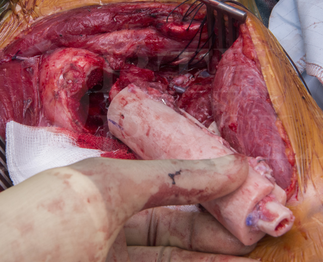
Learn the Intercalary humeral excision and allograft reconstruction with vascularised fibula (the Capanna technique) surgical technique with step by step instructions on OrthOracle. Our e-learning platform contains high resolution images and a certified CME of the Intercalary humeral excision and allograft reconstruction with vascularised fibula (the Capanna technique) surgical procedure.
Enchondromas are intramedullary neoplasms made of well-differentiated hyaline cartilage. The exact incidence is unknown as the majority are asymptomatic and discovered incidentally. Enchondromas only occur in bones that pre-formed in cartilage. The commonest location is the tubular bones of the hand followed by the femur and humerus. Radiographically, enchondromas can be very large with consequent expansion of the bone, and thinning or complete loss of the cortex.
Multiple enchondromas are rare. In the case of Ollier’s disease, multiple enchondromas may be found within the hand of one limb, or have a much wider, hemisomic distribution, or affect the entire body with a hemisomic prevalence. The disease is non-hereditary and sporadic. It most commonly affects the tubular bones of the hand or foot. In the case of Maffucci syndrome, multiple enchondromas are associated with multiple cutaneous or deep haemangiomas; the presence of haemangiomas may be identified radiographically as phleboliths. Histologically, the enchondromas appear more cellular than solitary enchondromas, with more proliferative histological potential.
Transformation to a secondary chondrosarcoma is seen in both these conditions. In Ollier’s, this may occur in 20-40% of patients whilst in Maffucci, this is much more common and is likely to be greater than 50%. Malignant transformation may be heralded by an increase in size of a lesion or the development of symptoms, typically pain. Both conditions are associated with an increased risk of extra skeletal malignancies such as breast, liver, ovarian and CNS tumours.
The indications for imaging and biopsy of enchondromas in the context of Ollier’s disease is the clinical suggestion of malignant transformation i.e. pain or increase in size. In this case the patient had previously sustained a pathological fracture with resultant deformity and continued to have painful symptoms and dysfunction after fracture union. A biopsy described features consistent with a benign cartilage neoplasm, but recent evidence has questioned the validity of pre-operative biopsy in determining grade in cartilage tumours (https://online.boneandjoint.org.uk/doi/abs/10.1302/0301-620X.100B5.BJJ-2017-1243.R1).
Author: Jonathan Stevenson
Institution: Royal Orthopaedic Hospital, Birmingham
Clinicians should seek clarification on whether any implant demonstrated is licensed for use in their own country.
In the USA contact: fda.gov
In the UK contact: gov.uk
In the EU contact: ema.europa.eu