Subscribe to get full access to this operation and the extensive Knee Surgery Atlas.
FREE TRIAL
Learn the Revision total Knee replacement: Second-stage with distal femoral EPR (Implantcast MUTARS MK) and EPORE collar and tibial cone surgical technique with step by step instructions on OrthOracle. Our e-learning platform contains high resolution images and a certified CME of the Revision total Knee replacement: Second-stage with distal femoral EPR (Implantcast MUTARS MK) and EPORE collar and tibial cone surgical procedure.
Increasing numbers of primary and revision knee replacements are being performed which inevitably lead to more prosthetic joint infections (PJI) presenting to specialist PJI multi-disciplinary teams. Infection is a devastating complication of total joint arthroplasty and not only the most common cause for early failure of joint replacements but also the most common cause for failure of revision knee replacements at any time.
That PJI is associated with higher mortality than some common malignancies has been widely reported. The five year survival after PJI is 78% compared to 90% in patients undergoing aseptic revision arthroplasty (Matar H, et al. Septic Revision Total Knee Arthroplasty Is Associated With Significantly Higher Mortality Than Aseptic Revisions: Long-Term Single-Center Study (1254 Patients). Journal of Arthroplasty 2021 https://doi.org/10.1016/j.arth.2021.01.068).
The treatment of prosthetic joint infection typically requires surgery involving explant of the infected prosthesis, radical debridement and then either immediate reimplantation or use of an antibiotic loaded cement spacer and delayed reimplantation (a two-stage revision, as in this case). Alternative strategies include debridement and implant retention with modular exchange (indicated in acute PJI) and single-stage revision (considered in infected primary arthroplasty implants, sensitive organisms and without soft-tissue defects requiring plastic surgery). There is endless debate about selecting the correct option for each case, the decision is multi-factorial and probably best decided in specialist PJI multi-disciplinary meetings.
Infected revision knee replacements are increasing in prevalence due to the increasing numbers of revision joint replacements being performed for septic and aseptic indications such that a 7.5-fold increase in re-revision knee replacements due to infection that has been experienced in the UK since 2005 (Lenguerrand et al. Description of the rates, trends and surgical burden associated with revision for prosthetic joint infection following primary and revision knee replacements in England and Wales: an analysis of the National Joint Registry for England, Wales, Northern Ireland and the Isle of Man. BMJ Open 2017;7:e014056. doi: 10.1136/bmjopen-2016-014056).
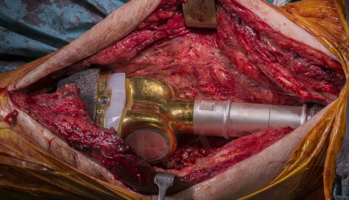
Dependent upon the medical condition of the patient, repeat two-stage revision for re-infected knee replacements has proven to be possible, although in immunocompromised hosts with poor soft-tissues, amputation should be considered. These limb-salvage procedures are challenging for numerous reasons. Orthopaedically dealing with segmental bone loss and poor residual bone stock means the choice of prosthesis is crucial and the Implantcast modular limb-salvage system has anti-infective silver, porous EPORE cones and EPORE collar and significant on-table flexibility which helps overcome some of these reconstructive challenges.
The outcomes however of repeat two-stage revision are not widely described; our own hospital data suggests that failure due to recurrent infection after 2 years can be as high as 50%, compared to 10% in primary two-stage revisions of infected primary knee replacements, so proceeding must be a carefully considered and consented intervention. Failure to control PJI may lead to further surgery, antibiotic suppression or amputation.
Here I present a challenging case of reconstruction of an infected revision knee replacement, which underwent a two-stage revision using a porous tibial cone for severe bone loss and a silver-coated endoprosthetic replacement to manage segmental bone loss.
Readers will also find the following related OrthOracle surgical techniques of interest:
First Stage Revision Total Knee Replacement for Acute Prosthetic Joint Infection (Zimmer Biomet articulating spacer)
Second Stage Revision Total Knee Replacement. PFC / MBT TKA with metaphyseal sleeve and stem (Depuy)
Revision total Knee Replacement: Legion CCK (Smith and Nephew)
Revision total Knee Replacement- Legion Rotating Hinge Knee ( Smith and Nephew)
Author: Jonathan Stevenson FRCS (Tr & Orth)
Institution: Royal Orthopaedic Hospital, Birmingham, UK
Clinicians should seek clarification on whether any implant demonstrated is licensed for use in their own country.
In the USA contact: fda.gov
In the UK contact: gov.uk
In the EU contact: ema.europa.eu