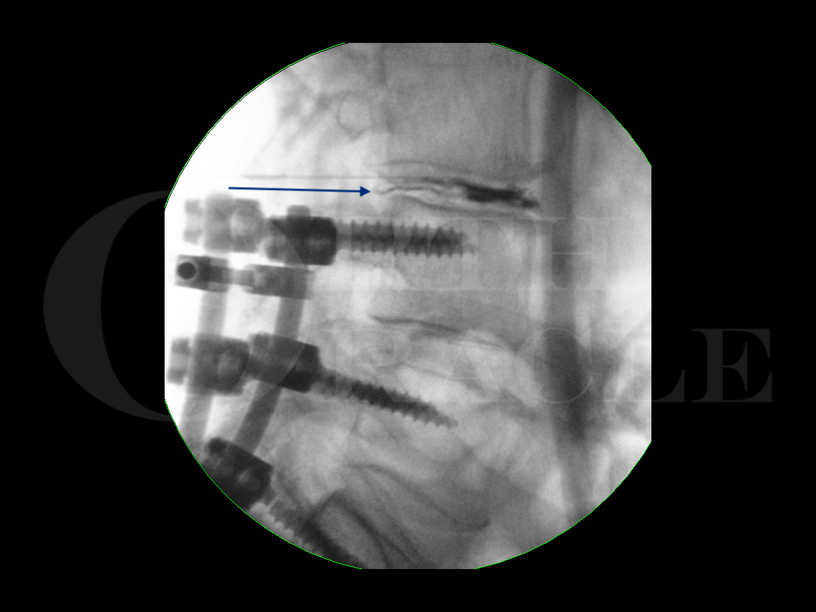
Lumbar discography (L3/L4)
Overview

Subscribe to get full access to this operation and the extensive Spine Surgery Atlas.
Learn the Lumbar discography (L3/L4) surgical technique with step by step instructions on OrthOracle. Our e-learning platform contains high resolution images and a certified CME of the Lumbar discography (L3/L4) surgical procedure.
The lumbar intervertebral disc has been viewed as a potential source for pain for many years. In 1934 Mixtel and Barr published their article on the intervertebral disc and its role in sciatica and since then the disc has been put forward as playing a role in discogenic back pain. The postulated mechanisms of action include stretching the fibres of the annulus fibrosus, extravasation of noxious chemicals from the disc around the nerve and dura, pressure on the nerves, granulation tissue within the annulus, and abnormal loading on the annulus or nucleus.
Lumbar discography is aimed at assessing the state of the disc and its part in the causation of a patient’s pain. X-rays, CT scans, and MRI scan are all imaging modalities that tell us about the anatomy of the disc but don’t directly indicate whether the disc is causing a patient’s pain.
Lumbar discography is not universally accepted as being an effective management due to the risk of false positives and complications such as discitis. Its use is therefore controversial.
The aim is to place the needle tip into the nucleus pulposus and then inject dye to assess the disc morphology, provoke pain by increasing the intradiscal pressure, and try to reduce the pain over time using local anaesthetic directly into the disc space. The provocative element of this test can be controlled using an adjacent disc space for comparison but I prefer to only inject into the disc space of interest due to the potentially devastating consequences of discitis in an otherwise normal disc space.
Fluoroscopy is required in multiple planes to navigate the needle tip safely into position within the disc space. A good working understanding of the local anatomy is essential especially with regard to Kambins Triangle. This is covered in the Nerve root block technique on OrthOracle at https://www.orthoracle.com/library/lumbar-nerve-root-block/. This is the posterolateral extradural approach which avoids the thecal sac. At the L3/4 level described here the needle trajectory enters the disc space low in Kambins triangle which is delineated medially by the dural edge, inferiorly by the superior endplate of the L4 vertebra below, and superolaterally by the L3 exiting nerve root.
I utilise a ‘needle inside needle’ technique which stops the inner needle (which will enter the intervertebral disc) from coming into contact with the skin. This is aimed at reducing the chance of discitis as a potentially devastating complication.
This technique describes lumbar discography, though the technique is also described in the thoracic and cervical spine. The complications in the cervical spine include epidural abscess, discitis, haematoma, and paralysis and given these has understandably limited indications.
The technique here deals with the L4/5 level. The L5/S1 level is possible but access is more difficult due to the narrow corridor of approach imposed by the iliac crest obstructing the trajectory.
Author: Mr Andrew Young, FRCS (Tr & Orth)
Institution: The RoyalOrthopaedic Hospital, Birmingham, UK.
Clinicians should seek clarification on whether any implant demonstrated is licensed for use in their own country.
In the USA contact: fda.gov
In the UK contact: gov.uk
In the EU contact: ema.europa.eu
Online learning is only available to subscribers.


