Subscribe to get full access to this operation and the extensive Spine Surgery Atlas.
FREE TRIAL
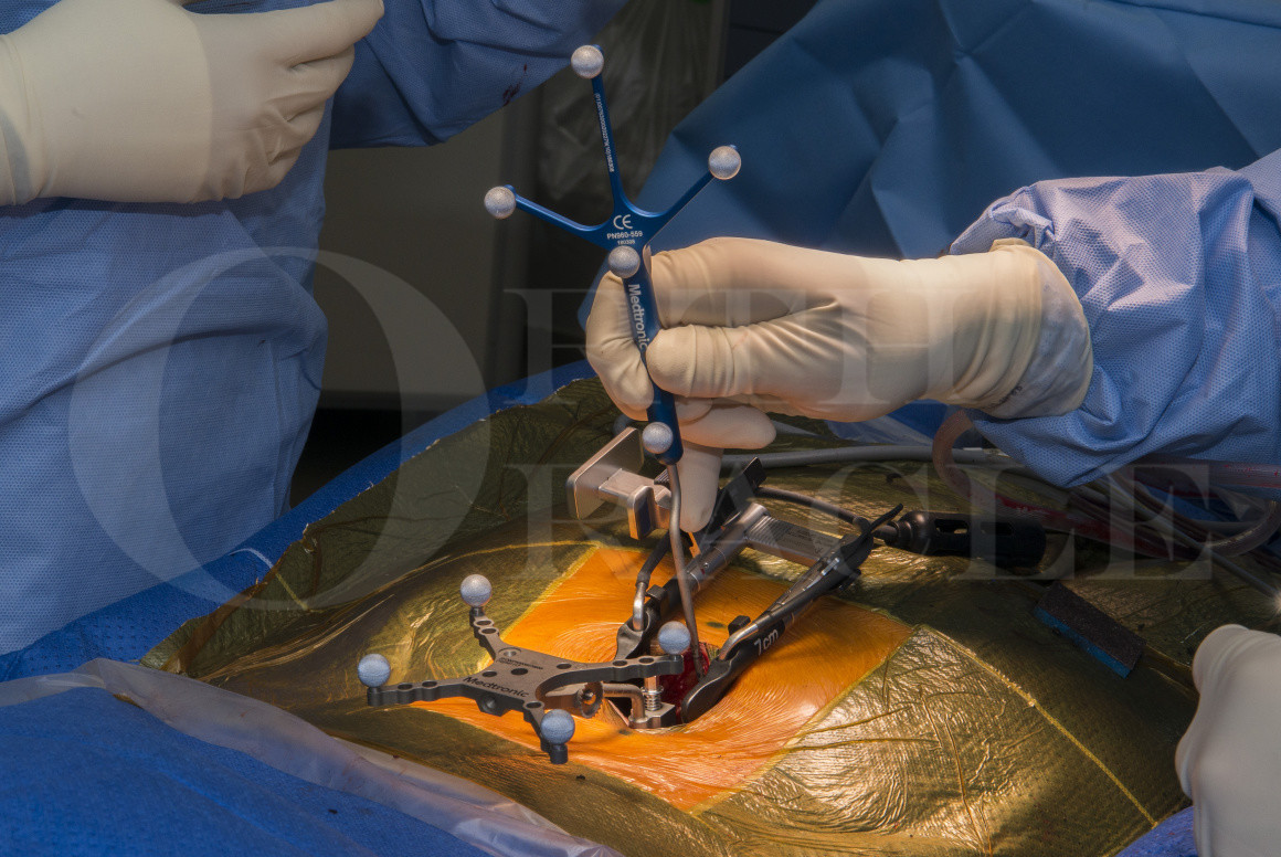
Learn the Minimally invasive posterior lumbar spine fusion (MidLIF) and decompression for spondylolisthesis with spinal stenosis (Medtronic Solera and Artic-L cage) surgical technique with step by step instructions on OrthOracle. Our e-learning platform contains high resolution images and a certified CME of the Minimally invasive posterior lumbar spine fusion (MidLIF) and decompression for spondylolisthesis with spinal stenosis (Medtronic Solera and Artic-L cage) surgical procedure.
Lumbar spinal stenosis is a very common condition affecting up to up to 47% of adults over 60 years old, although only around 9% may be symptomatic. Symptoms often come on with standing or walking and improve with leaning forwards or sitting down. In some instances, patients may also experience a radiculopathy while at rest.
Spinal stenosis can occur at any level of the spine. It is most commonly caused by facet joint hypertrophy, ligamentum flavum hypertrophy, and degenerative broad disc bulges. Since the lumbar spine and cervical spine move more than the thoracic spine, stenotic changes are more common in these mobile sections of the spine. More forces go through the lumbar spine which means stenotic changes are most common in this area.
Spinal stenosis can effect the central canal and lateral recesses but may also cause foraminal stenosis. It can also be associated with spondylolisthesis, retrolisthesis, and lateral listhesis as well as any spinal deformity. There is also a subgroup of patients who have congenital stenosis that predisposes them to becoming symptomatic if they develop degenerative changes later in life.
The condition often presents insidiously with spinal claudication. This manifests itself as paraesthesia or aching, progressing to pain and weakness. Patients typically have spinal claudication effecting both lower limbs but it can present unilaterally. The pain often starts when patients have been standing in one position for more than a few minutes, or if they walk for more than 5-10 minutes. Patients will often complain symptoms are worse if they have to walk slowly around shops. However, leaning forward on a stick, frame, or shopping trolley often alleviate their symptoms since flexion makes the lumbar spinal canal wider. Patients also describe that they need to sit down or lean forwards to allow their symptoms to resolve.
The vast majority of patients can be managed non-operatively and should be encouraged to stay as active as possible, with modification of activities that bring on their symptoms. However, for those who have exhausted non-operative measures, surgery may be beneficial.
Surgical management options available include lumbar decompression or fusion with decompression. In some patients, decompression procedure alone may not achieve satisfactory decompression of the nerves to improve symptoms. In this situation, the surgeon needs to work out if excision of the facet joints will achieve decompression. If the facet joints are excised, instrumentation is required to achieve stability of that spinal segment.
When performing a fusion operation, there are various options available to surgeons. These may involve posterior, anterior, or lateral approaches which can be open or minimally invasive.
The reason to consider a minimally invasive posterior fusion and decompression (MidLIF) is that satisfactory stability can be achieved with appropriate implants whilst resulting in less muscle stripping, which makes it easier for patients to recover than for open procedures. Biomechanical studies have found good pullout strength of the cortical bone trajectory screws used for this technique, including in osteoporotic bone. I have used Medtronic Solera 4.75 multiaxial screws which can be navigated using the O-arm; for lumbar degenerative work, it is uncommon to require fixed angle screw heads.
Many surgeons using this technique use a posterior interbody (PLIF) cage since the required access via the spinal canal is easier than a transforaminal approach. However, I use a transforaminal interbody (TLIF) cage since this can be positioned anteriorly in the disc space in order to create lordosis across the segment, compared to a (PLIF) cage which are often less lordotic and allow less compression posteriorly. The Medtronic Artic-L interbody cage used here has been designed with a hinged introducer which can be locked and unlocked to allow rotational control of the cage at all stages during insertion and this aids positioning it during minimally invasive procedures.
Author: Mr Stephen Morris FRCS (Tr & Orth).
Institution: The Avon Orthopaedic Centre, Southmead Hospital, Bristol, UK.
Clinicians should seek clarification on whether any implant demonstrated is licensed for use in their own country.
In the USA contact: fda.gov
In the UK contact: gov.uk
In the EU contact: ema.europa.eu