Learn the Calcaneal fracture: fixation with extended lateral approach and ZimmerBiomet ALPS plate surgical technique with step by step instructions on OrthOracle. Our e-learning platform contains high resolution images and a certified CME of the Calcaneal fracture: fixation with extended lateral approach and ZimmerBiomet ALPS plate surgical procedure.
Calcaneal fractures account for 1-2% of all fractures. They generally result from high energy mechanisms, most commonly falls from heights or road traffic accidents. The ‘rule of 10’ is useful when assessing these injuries, approximately 10% are bilateral, 10% open injuries and 10% associated with spinal injuries, usually thoracic-lumbar burst fractures.
They are in the main very significant injuries when intra-articular in nature, and require a clear understanding of the fracture anatomy, and patient factors (most importantly compliance and avoidance of smoking) as well as what can be realistically achieved with operative techniques, when forming an opinion and counselling patients on how to be treated.
Controversy surrounds their management, in particular whether open reduction and internal fixation is ever warranted, given a not inconsequential incidence of complications related to open approaches such as with the extended lateral approach described in some series. Such complications include wound breakdown, deep infection and pain syndromes related to cutaneous nerve compromise. This has led to the development of less invasive techniques to reconstruct calcaneal fractures including sinus tarsi approaches and percutaneous techniques.
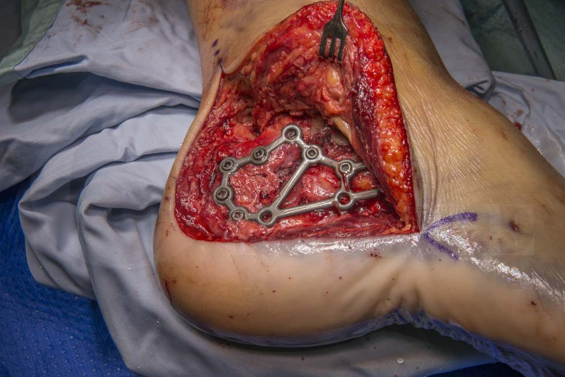
The extended lateral approach affords excellent visualisation of the components of the injury that require reduction, namely the lateral wall, subatalar and Calcaneo-cuboid joints and peroneal tendons. It is based one the angiosome of the lateral calcaneal artery, a terminal branch of the peroneal artery. The use of the extended lateral approach was popularised by, amongst others, Professor Roger Atkins at The Bristol Royal Infirmary, UK. His two seminal papers from 1993, published in the British Journal of Bone and Joint Surgery (and available open access), describe the patho-anatomy, surgical approach and sequence of fixation. They are essential reading for anyone embarking on the surgical fixation of articular depression fractures of the calcaneum, regardless of approach used.
The UK heel fracture and its accompanying headline in the BMJ caused significant controversy with its assertion that open reduction and internal fixation should not be recommended for displaced intra-articular fractures. The debate over this paper continues but it is certainly true that newer techniques and implants continue to develop which avoid the need for the use of the extended lateral approach. It is also true that these fractures should be managed by surgeons and units used to dealing with large volumes of these injuries- the median number of operations per surgeon in the study was 2 and this may be related to the high rate of complications specifically a 19% infection rate which any surgeon or unit would deem unacceptable. As with many aspects of complex trauma, rather than didactically deciding on treatment based on a simple radiological review, the decision making as to the best treatment for an individual patient relies on careful examination of the injured limb, study of the X-rays and scans and a detailed discussion with the patient as to the risks and benefits of each treatment for them in light of other factors such as smoking or medical comorbidities- unfortunately this nuanced process does not lend itself to an RCT.
In this case I have used the Zimmer-Biomet ALPS plating system. I prefer these implants when fixing calcaneal fractures with an extended lateral approach. The instrumentation is well designed, there are good screw options including cortical, locking and variable angle screws and crucially the plate and screws have a very low profile once the fixation is completed minimising the risk of soft tissue irritation or impingement.
Readers will also find the following OrthOracle operative techniques of interest:

 00:00
00:00


