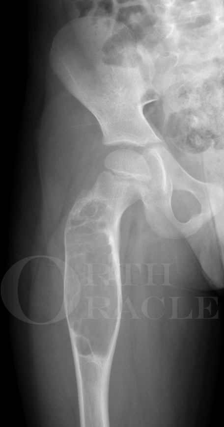
Unicameral bone cyst (proximal femur) : curettage, bone grafting and plating
Overview

Subscribe to get full access to this operation and the extensive Bone & Soft Tissue Tumour Surgery Atlas.
Learn the Unicameral bone cyst (proximal femur) : curettage, bone grafting and plating surgical technique with step by step instructions on OrthOracle. Our e-learning platform contains high resolution images and a certified CME of the Unicameral bone cyst (proximal femur) : curettage, bone grafting and plating surgical procedure.
Simple or unicameral bone cysts are common benign lesions found in children and adolescents and were first described by Virchow in 1891. It is important to distinguish them from more aggressive lesions such as aneurysmal bone cysts. Simple bone cysts (SBC) will frequently heal spontaneously once the adjacent physis has closed at skeletal maturity but lesions are still observed in some adults.
The majority of lesions are found in the metaphysis of long bones, the humerus being the most frequent site. However, any bone can be effected including the axial skeleton. Boys are more frequently affected than girls.
Simple bone cysts (SBC) are frequently asymptomatic unless they result in pathological fracture. The effects of such fractures is largely determined by their location, one of the most significant being around the hip where such injuries can result in malunion and damage to the blood supply to the femoral head unless appropriately managed.
Simple bone cysts can be treated by several modalities. More aggressive management is generally indicated for the proximal femur because of the consequences of femoral neck fracture.
Author: Christopher Edward Bache FRCS (Tr & Orth)
Institution: The Royal Orthopaedic Hospital, Birmingham, UK.
Clinicians should seek clarification on whether any implant demonstrated is licensed for use in their own country.
In the USA contact: fda.gov
In the UK contact: gov.uk
In the EU contact: ema.europa.eu
Online learning is only available to subscribers.


