Subscribe to get full access to this operation and the extensive Shoulder & Elbow Surgery Atlas.
FREE TRIAL
Learn the Elbow arthroscopy surgical technique with step by step instructions on OrthOracle. Our e-learning platform contains high resolution images and a certified CME of the Elbow arthroscopy surgical procedure.
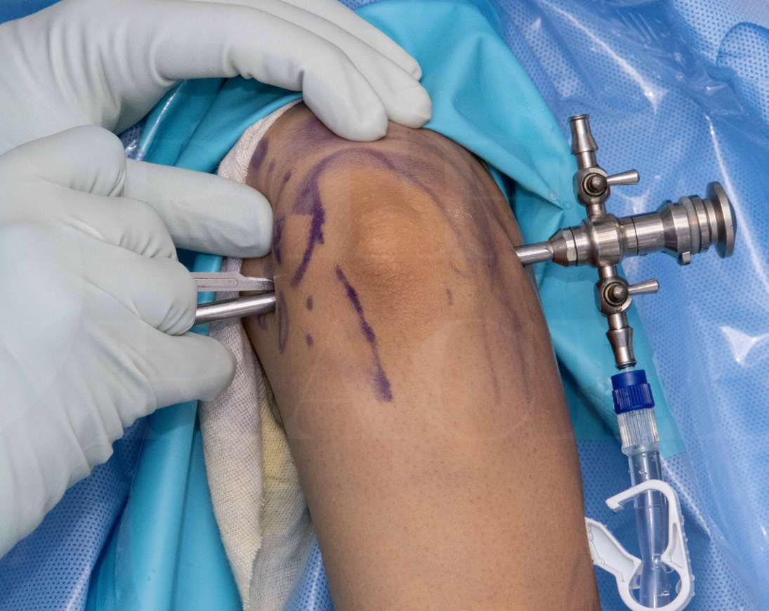
Arthroscopy of the elbow can be conducted both for diagnostic purposes and to undertake therapeutic procedures. Although there is one single joint cavity, the elbow comprises articulations between the humerus and both the ulna (ulno-trochlear) radius (radio-capitellar) and additionally between the two forearm bones (the proximal radioulnar joint). The ulno-humeral articulation also has a posterior compartment, and so for a complete even diagnostic arthroscopy, multiple portals will be needed.
The placement of these portals will need to bear in mind the position of important neurovascular structures that pass close to the elbow, and unlike in the knee and the shoulder, these will be found on both sides of the joint; the positioning and technique of creating the various arthroscopic portals will therefore be influenced both by the access required to complete the surgical task, and also a secure knowledge of the anatomy of the structures surrounding the elbow, including potential variations. However, as long as these factors are considered, arthroscopy of the elbow can be a very rewarding and enjoyable undertaking.
Usually, arthroscopy of the elbow will be undertaken with the patient in a lateral decubitus position, the body supported securely and with the arm supported with a arm holder, or resting across a padded bar. Traction on the elbow is not necessary as the joint cavity will permit visualisation without distraction being applied. For most cases, a standard 4mm 30° side-viewing arthroscope arthroscope can be used for elbow arthroscopy, although in very small individuals (children and small adolescents) a 2.9mm wrist or ankle arthroscope may be preferred.
OrthOracle readers will also find the following arthroscopic instructional techniques of interest:
Diagnostic Wrist Arthroscopy (using Acumed ARC Tower )
Ankle arthroscopy using the Smith and Nephew Guhl non-invasive ankle distractor
Shoulder arthroscopy: Arthroscopic capsular release and MUA
Hip Arthroscopy: set up and access to central compartment
Subtalar fusion: Arthroscopic technique
Diagnostic knee arthroscopy
Author: Chris Little FRCS (Tr & Orth)
Institution: The Nuffield Orthopaedic Centre, Oxford, UK.
Clinicians should seek clarification on whether any implant demonstrated is licensed for use in their own country.
In the USA contact: fda.gov
In the UK contact: gov.uk
In the EU contact: ema.europa.eu