Learn the Sural nerve graft reconstruction of the sciatic nerve after resection of a malignant peripheral nerve sheath tumour surgical technique with step by step instructions on OrthOracle. Our e-learning platform contains high resolution images and a certified CME of the Sural nerve graft reconstruction of the sciatic nerve after resection of a malignant peripheral nerve sheath tumour surgical procedure.
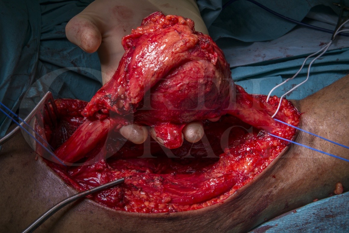
Autologous reversed sural nerve cable grafting is the gold standard method for reconstruction of large nerve gaps in mixed (motor and sensory) major nerve trunks. Functional recovery is determined by the gap length, the site of the reconstruction, the duration of denervation, the surgical graft bed, the quality of the target muscles and the age of the patient. Malignant peripheral nerve sheath tumours (MPNST) are rare and usually present with a rapidly enlarging and painful mass with associated sensory and motor deficits. There may be a history of a neurofibromatosis which is associated with malignant transformation in peripheral nerve sheath tumours. Magnetic resonance imaging and biopsy are used to determine the diagnosis. The pathophysiological grade and the stage of disease determine the prognosis. Staging computed tomography will identify whether there are any metastases at presentation.
The surgical management involves excision of the tumour with a cuff of normal tissue to achieve an adequate surgical margin. Whilst the exact width of an adequate margin remains a sourc of constant debate, it should take into consideration the histological subtype, the quality of the margin tissue, the proximity of nearby vital structures and the use of adjuvant or neo adjuvant radiotherapy. In the case of large tumours and high grade histologies, compartment excision or ablative surgery may need to be considered to achieve an adequate margin. When post-operative radiation is required, non-vascularised autologous graft reconstruction is unlikely to support useful neural regeneration. In lower grade tumours, gap reconstruction with autologous graft provides a scaffold for nerve regeneration and results in less neuropathic pain, some proximal motor recovery and the chance of protective sensation to the plantar surface of the foot. Biopsy may lead to sampling error and the final grading of the tumour and adequacy of excision can only be determined on final specimen histopathological examination.
The case presented is a Malignant peripheral nerve sheath tumours that was biopsied and reported as low grade and surgical excise and bilateral autologous rural nerve cable grafting performed for gap reconstruction. The technique presented here will focus on the grafting technique and alternative options.
Institution: Peripheral Nerve Injury Service & Royal Orthopaedic Hospital, Birmingham, UK
Surgical Steps
Welcome to the surgical steps quiz.
This tests your ability to recall the correct order in which the key steps of an operation should be performed.
There will be various legitimate variations in this order but the quiz doesn’t reflect this.
Your reference point for the steps quiz is just our operative technique.
It’s one starting point in helping you to learn the technique.
You simply “drag & drop” the boxes containing the descriptions of the steps into the order you think fits and submit for marking.
If you get it correct first time you will be presented your certificate at the end of the quiz.
If you need to do more work on the steps you access your marked version by clicking the“re-sit the quiz” button.
This will show the steps you ordered correctly highlighted in green and the incorrect ones in red.
The correct green steps are fixed and won’t move.
The red steps move differently after your initial attempt and swap locations with the step they are dragged and dropped onto .
Finally resubmit the quiz once again.
The quiz will let you keep going until you get all the steps in the right order.
The correct green steps are fixed and won’t move.
The red steps move differently after your initial attempt and swap locations with the step they are dragged and dropped onto .
Finally resubmit the quiz once again.
The quiz will let you keep going until you get all the steps in the right order.
COURSE PROGRESS
surgical steps
Time limit: 0
Quiz Summary
0 of 1 questions completed
Questions:
Information
You have already completed the quiz before. Hence you can not start it again.
Quiz is loading…
You must sign in or sign up to start the quiz.
You must first complete the following:
Results
Quiz complete. Results are being recorded.
Results
0 of 1 questions answered correctly
Your time:
Time has elapsed
Categories
Not categorized0%
1
Current
Review
Answered
Correct
Incorrect
Question 1 of 1
1. Question
The cables are positioned to cover the proximal nerve stump and built up progressively.
The deep fascia is opened and the interval between the medial and lateral hamstring muscles is identified
The sural nerve is identified midway between the posterior aspect of the lateral malleolus and the achilles tendon at the level of the ankle.
The sural nerve is inserted as a series of cables across the nerve gap connecting fascicle groups.
The resected tumour is tagged with orientation sutures to enable the histopathologist to define adequacy of resection margins.
The sural graft donor length is maximised on the right leg prior to sectioning and removal of the graft.
Next the surgical team change gown and gloves and open new instrumentation for the next phase of the surgery which involves opening sural nerve donor sites in both lower legs
Tisseel tissue glue is applied and produces a fibrin clot around the coaptation sites and between the fascicles, providing support and resistance to disruption of the nerve-graft interface.
At the distal end of the tumour the dissection in the popliteal fossa identifies the tibial nerve, the common peroneal nerve and the popliteal vessels.
The patient is placed prone under general anaesthesia on a Montreal mattress with the left thigh tumour site marked.
A Norfolk and Norwich retractor is placed in the interval between the medial and lateral hamstring muscles.
The skin is incised and the posterior cutaneous nerve of the thighs' terminal branches may be seen and should be protected
The distal neurorraphies are sutured and then Tisseel glue is used to provide additional support, as for the proximal neurorraphy.
The muscle layer is opposed over the graft and secured with interrupted absorbable vicryl sutures.
The distal tibial nerve and common peroneal nerves are also sectioned with a scalpel blade.
Both lower limbs are prepped and draped.
The sural nerve is mobilised away form the short saphenous vein using the sloop to minimise handling of the nerve during harvest.
Once the sural nerve is identified the skin can be incised along the expected course of the nerve.
Careful coagulation with bipolar diathermy to the cut sciatic nerve face
The assistant continues with the right leg sural nerve harvest under tourniquet. The main surgeon exposes the sural nerve on the left leg which has had tumour resection.
Dissection continues to separate the nerve from the short saphenous vein.
The upper sciatic nerve is sectioned at least 5cm above the macroscopic tumour extent.
The sciatic nerve is identified in the upper wound above the tumour.