Subscribe to get full access to this operation and the extensive Foot Surgery Atlas.
FREE TRIAL
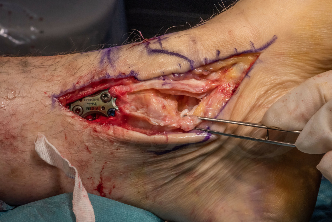
Learn the Ankle instability (Medial): Deltoid ligament and tibialis posterior sheath repair surgical technique with step by step instructions on OrthOracle. Our e-learning platform contains high resolution images and a certified CME of the Ankle instability (Medial): Deltoid ligament and tibialis posterior sheath repair surgical procedure.
The indications for surgery to the deltoid ligament can be considered in terms of the acute injury setting, where there is a degree of controversy about the necessity for deltoid ligament repair, and the chronic setting in the presence of longstanding planovalgus deformity and/or medial ankle instability where there is little controversy about repair of the structure. These are discussed in more detail in the indications section.
The deltoid ligament is composed of superficial and deep components which are confluent with each other. The superficial part of the deltoid is the main restraint to ankle and hindfoot valgus and the deep component resists external rotation. Anatomically, the superficial deltoid runs from the medial malleolus as a fan shaped structure its’ described components being the tibionavicular, tibiospring, tibiocalcaneal and plantar calcaneonavicular. The deep component of the deltoid also takes origin from the tip of the medial malleolus, though inserts in two bands, both into the talus, the anterior tibiotalar and the posterior tibiotalar.
Medial deltoid ligament reconstruction is notorious for its difficulty primarily due to its complex anatomical structure and the difficulties in being able to tension the ligament correctly. Re-ruptures in chronic cases are not infrequent because, suturing of degenerate ligament is difficult, as is the ability to securely attach a thickened and myxoid structure back on to the medial malleolus.
Readers should also familiarise themselves with Kartik Hariharans’ excellent technique :
Supra-malleolar distal tibial osteotomy: Medial closing wedge, deltoid ligament repair and ankle arthroscopy.
This contains a very detailed series of images and instructional steps explaining reconstruction of the deltoid ligament using a bone tunnel technique with the Arthrex internal brace implant.
Readers will also find the following associated techniques of interest :
Pes planus correction : Calcaneal osteotomy(Wright DARCO calcaneal plate), spring ligament reconstruction and Flexor digitorum longus transfer.
Pes Planus correction: Lateral column lengthening and medial column fusion ( over-corrected club foot deformity)
Pes Planus correction with FDL transfer, Calcaneal osteotomy and Wright Bioarch arthroresis screw
Tibialis Posterior reconstruction for pes planus, using FDL transfer and Arthrex Biotenodesis screw.
Lateral ankle ligament reconstruction: Broström (mid-substance) repair
Lateral ankle ligament reconstruction: Ligament advancement using twin fix suture anchors(Smith and Nephew)
Lateral ankle ligament reconstruction: Brostrom technique
(Thanks to Peter Rosenfeld FRCS Tr & Orth for the images of his case which are shown in this technique).
Author: Mark Herron FRCS
Institution: OrthOracle, London, UK.
Clinicians should seek clarification on whether any implant demonstrated is licensed for use in their own country.
In the USA contact: fda.gov
In the UK contact: gov.uk
In the EU contact: ema.europa.eu

 00:00
00:00


