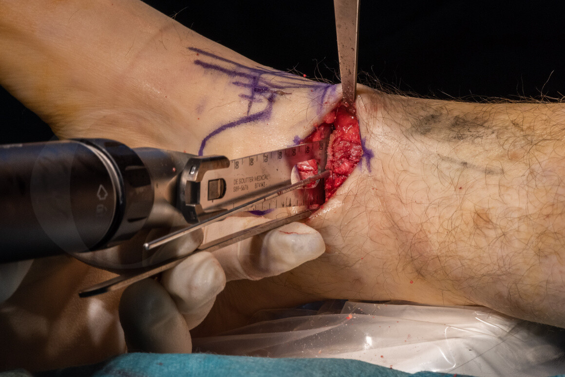
Supra-malleolar distal tibial osteotomy: Medial closing wedge, deltoid ligament repair and ankle arthroscopy
Overview

Subscribe to get full access to this operation and the extensive Foot Surgery Atlas.
Learn the Supra-malleolar distal tibial osteotomy: Medial closing wedge, deltoid ligament repair and ankle arthroscopy surgical technique with step by step instructions on OrthOracle. Our e-learning platform contains high resolution images and a certified CME of the Supra-malleolar distal tibial osteotomy: Medial closing wedge, deltoid ligament repair and ankle arthroscopy surgical procedure.
Ankle arthritis in a majority of patients is post traumatic, either as a result of intra-articular ankle fracture, osteochondral injury, malalignment of the tibia following fracture, ligamentous injuries including lateral and medial collateral ligaments and less commonly, significant syndesmotic instability. Aside from inflammatory arthritides, trauma is the main reason for the development of osteoarthritis of the ankle. Rarer causes are secondary to previous episodes of avascular necrosis, sepsis or chondrolysis. Idiopathic ankle arthritis does occur (where no specific cause is identifiable) but is far rarer than with the knee or hip where it is the commonest group.
Ankle arthrodesis has been the mainstay of treatment for many of these conditions when they have become sufficiently symptomatic and conservative measures have failed. However, over two decades ago, it was recognised that asymmetric arthritis (non-concentric arthritis) was a significant entity in patients who have either an extra-articular supramalleolar deformity resulting in malalignment of the axis of weight bearing, or have ligamentous instability leading to malpositioning of the talus within the ankle mortise. The latter group of patients often had rotational malalignment of the talus within the mortise, due to the rotatory subluxation that is caused by significant ligamentous deficiency in the medial or lateral aspect. Thus, for example, medial ligament deficiency (deltoid deficiency), results in an external rotation valgus deformity, with the talus lying in this abnormal orientation within the ankle mortise. This, over a period of time, results in asymmetric arthritis of the lateral part of the ankle joint, including the lateral gutter. Similarly, patients with lateral ligament deficiency often present with asymmetric varus deformities, including the medial gutter, with internal rotation also forming a component.
The supramalleolar osteotomy is a joint sparing surgical technique for the management of asymmetric symptomatic early arthritis of the ankle. In recent years, surgeons like Hintermann, Takakura, and Myerson have expanded the technique and indications for a supramalleolar osteotomy for addressing asymmetric arthritis of the ankle joint. Significant difficulties have been encountered in the indications for this procedure and the use of additional procedures continuing to be researched as thus far, they still appear to be somewhat arbitrary. Attempts are being made to differentiate types of asymmetric arthritis into several sub-sections and based on these subdivisions various classifications have been propounded. These groups are used in determining the choice of procedure, as well as also identifying potential negative predictors of the outcome of this operation.
Moreover, additional underlying foot deformities, severity of deformity, difficulty in accurately mapping the deformity, and in particular the rotational component of the deformity, have caused some concerns about utilising this procedure for the treatment of early to mid-range arthritic change, which occurs in the ankle in an asymmetric fashion. Finally, the limit to which supramalleolar osteotomy can be used with predictable results, as a treatment option which lies between ankle arthrodesis and total ankle replacement, has not been clearly established, which emphasises the importance of patient education and involvement in the decision making process.
A medial closing wedge osteotomy is utilised in patients with asymmetric valgus arthritis of the ankle. This may have a component of external rotation deformity and is classically caused with deltoid ligament deficiency, resulting in this deformity.
The resultant axis of load transfer through the foot and the ankle into the tibia is therefore lateralised as a result of the deformity, where the hindfoot is now in valgus as a consequence of asymmetric wear in the lateral and posterior aspects of the joint. This often results in painful symptoms in the lateral aspect of the ankle selectively. The medial cartilage of the ankle is often noted to be normal on MRI scanning or arthroscopy. This is as a consequence of very little low transfer through the medial part of the articular surface of the talus and the tibia. Over a period of time, as this deformity worsens and there is eventual bone on bone contact, it is often the case that the talus seam starts to erode into the tibial articular surface and into subchondral regions of bone, resulting in lateral bone deficiency. This would render the ankle relatively unsuitable for a supramalleolar osteotomy, as this procedure does not address the lateral bone loss that is associated with a progressive deformity of this nature.
A supramalleolar osteotomy therefore is a method by which the access of low transfer is centralised and often medialised by excising a medial wedge that rotates the hind foot into a neutral position. This has the resultant effect of increasing load bearing on the normal cartilage on the medial side of the joint and decreasing the stresses that pass through the cartilage deficient aspect of the ankle on the lateral side. Furthermore, it also de-rotates the talus and into inversion with appropriate ligament repair, thereby decreasing the contact between the lateral surface of the talus and the articular surface of the lateral malleolus, thus decreasing painful stresses that are likely to go through this region in the deformed position.
It must be understood that there are limitations to what this procedure can achieve and in general, requires a combination of procedures with ligament repair and wedge excision in order to achieve optimal results. The long-term effects of this osteotomy are still being studied and there are no significant landmark publications that suggest that this operation may indeed reverse the process of progressive degenerative arthritis once the ankle is realigned. However, acquired knowledge from the high tibial osteotomies that are much more commonly used in the treatment of uni-compartmental knee arthritis, would suggest that realignment of the ankle may have similar results, as long as there is no osseous defect.
In this particular case, the three procedures of arthroscopy and microfracture, supramalleolar medial closing wedge osteotomy and a medial deltoid reconstruction were performed. The arthroscopic microfracture was to encourage fibrocartilaginous coverage of the lateral aspect of the talus, with the hope of producing secondary congruity, and the deltoid reconstruction providing the necessary rotational stability to the ankle, to prevent it from returning to its externally rotated valgus position.
In my experience of about a dozen or so cases over the last 10 years, I have found over 80% of patients were very satisfied with the outcome of the procedure, both in terms of pain relief, but also with regard to the functional improvement that this operation or combination of operations seems to achieve. About 20% of these patients eventually went on to an ankle replacement or an ankle fusion. The supramalleolar osteotomy can also be regarded as an attractive as an interim term solution, prolonging the functional life of these ankles, whilst making subsequent fusion or ankle replacement much easier as the ankles are well aligned for these procedures as a result of the supramalleolar osteotomy.
The plate I have used in this operation is the Arthrex H-plate without a wedge. These are generally used for lateral column lengthening, calcaneo-cuboid fusions and other procedures that benefit from low profile, compression and locking screw options. The plate is of a low profile, can be contoured, and can be used both in static or compression mode. They come in left and right slants with the inner holes being compression holes and the outer being locking (both fixed and variable locking angles) holes. The compression option is utilised by drilling eccentrically in the compression holes.
OrthOracle readers will also find the following associated instructional surgical techniques of interest:
Supra-malleolar distal tibial osteotomy: Medial opening wedge with Arthrex plate.
Supra-malleolar distal tibial osteotomy: Minimally invasive technique with the Taylor Spatial Frame
Ankle arthrodesis(fusion): Mini-open technique (Arthrex 6.7mm Titanium cannulated screws)
Ankle Arthrodesis (Fusion): Using Arthrex anterior ankle fusion plate
Ankle Arthrodesis(Fusion): Trans-fibular approach using AnkleFix 4.0 plate (Zimmer-Biomet)
Ankle arthrodesis (fusion): Arthroscopic assisted Ankle Fusion
Ankle arthroscopy using the Smith and Nephew Guhl non-invasive ankle distractor
Ankle arthrodesis (fusion): Trans-fibular approach
There are also numerous ankle replacement techniques on OrthOracle that will be of interest to readers.
Author: Kartik Hariharan FRCS.
Institution: Aneuran Bevan University Health Board, Wales.
Clinicians should seek clarification on whether any implant demonstrated is licensed for use in their own country.
In the USA contact: fda.gov
In the UK contact: gov.uk
In the EU contact: ema.europa.eu


