Subscribe to get full access to this operation and the extensive Hip Surgery Atlas.
FREE TRIAL
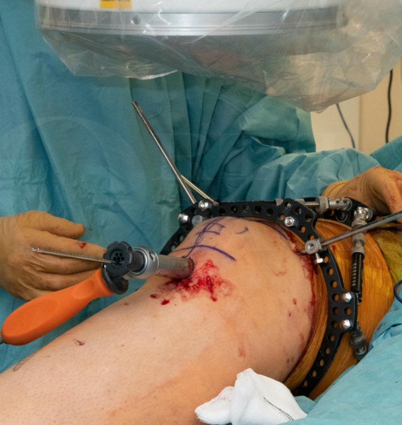
Learn the Femoral deformity correction: CHAOS technique using Taylor Spatial Frame and Trigen Nail (Smith and Nephew) surgical technique with step by step instructions on OrthOracle. Our e-learning platform contains high resolution images and a certified CME of the Femoral deformity correction: CHAOS technique using Taylor Spatial Frame and Trigen Nail (Smith and Nephew) surgical procedure.
Long bone deformity in the lower limb may follow trauma, be acquired secondary to metabolic bone disease or may be the result of congenital deformities. Deformity in the long bones which results in a mechanical axis deviation can, in the longer term, result in arthritic changes in both the knee and the ankle if untreated. The magnitude of deformity which will result in longer term joint damage is however controversial with no universally accepted figures. Probably deformities up to 5 degrees in the coronal plane and 10 degrees in the sagittal plane are tolerated however this will depend on the location of the deformity in the affected bone.
When deformity correction is considered careful planning is required. This has been outlined in the ‘Introduction to the taylor spatial frame and deformity planning’ case. The first step involves plotting the mechanical axis of the limb from the centre of the hip to the centre of the knee to determine whether a varus or valgus deformity of the leg is present. Next, one identifies which segment of the limb is involved by plotting the joint line angles around the knee and comparing these either with the normal contralateral side if no to deformity is present theret or using published population normal values. This one to identify whether the deformity lies within the tibia or the femur or both bones. Once the involved bone has been identified one locates the position of the deformity in the bone by plotting the axis of the proximal and distal segments of the bone to identify the CORA (centre of rotation of angulation). With this information we are now able to assess the magnitude of the deformity, to plan where to perform an osteotomy to allow the deformity to correct and in addition plan where to place either a physical or virtual hinge to perform the correction and then to decide how to stabilise the osteotomy during and after correction.
Deformity correction can be performed acutely, when the soft tissues will allow this, or gradually in which case the soft tissues have time to adapt and correct also. If the deformity is corrected acutely then stabilisation with internal fixation using either an intermediary nail or plate may be appropriate. In the majority of cases of gradual correction a ring fixator is the preferred method of stabilisation. A further benefit of using a ring fixator is that it will allow fine tuning of the correction post-operatively and a more accurate final correction then with acute correction and internal fixation.
Due to the challenges of acutely correcting complex deformities, particularly when in more than one plane, using standard internal fixation techniques various methods have developed to combine the benefits of more accurate correction with external fixator together with the use of internal fixation to stabilise the final correction and thereby avoiding the need for prolonged time in an external fixator for the patient. One such method is the computer hexapod assisted orthopaedic surgery (CHAOS) technique as described by the Bristol Limb Reconstruction unit (see reference below). This method uses a hexapod frame to perform complex deformity correction acutely in theatre. Once the correction has been completed the osteotomy and is stabilised with internal fixation and the frame is then removed. This technique allows accurate deformity correction without the need for prolonged postoperative time in a ring fixator.
Hughes A, Heidari N, Mitchell S, Livingstone J, Jackson M, Atkins R, Monsell F. Computer hexapod-assisted orthopaedic surgery provides a predictable and safe method of femoral deformity correction. Bone Joint J . 2017 Feb;99-B(2):283-288.
Readers will also find the following associated OrthOracle techniques of interest:
Taylor Spatial Frame(Smith and Nephew). Introduction to hardware, frame application and use of software for deformity correction.
Tibial fracture non-union correction using Taylor Spatial Frame (Smith and Nephew)
Tibial shaft fracture: Fixation with a Taylor Spatial Frame (TSF) circular external fixator (Smith and Nephew)
Supra-malleolar distal tibial osteotomy: Minimally invasive technique with the Taylor Spatial Frame
Limb reconstruction
Author: Mr Paul Fenton FRCS (Tr & Orth)
Institution: The Queen Elizabeth Hospital, Birmingham, UK.
Clinicians should seek clarification on whether any implant demonstrated is licensed for use in their own country.
In the USA contact: fda.gov
In the UK contact: gov.uk
In the EU contact: ema.europa.eu