Subscribe to get full access to this operation and the extensive Hip Surgery Atlas.
FREE TRIAL
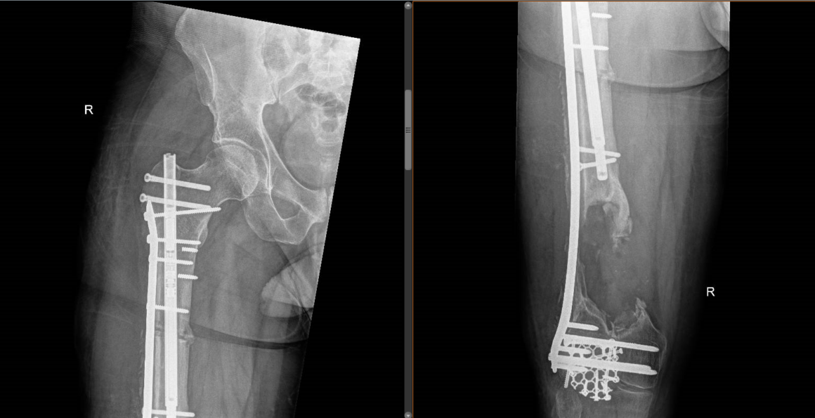
Learn the Femoral shaft fracture: Plate assisted bone transport using Precice Bone transport nail (Nuvasive) surgical technique with step by step instructions on OrthOracle. Our e-learning platform contains high resolution images and a certified CME of the Femoral shaft fracture: Plate assisted bone transport using Precice Bone transport nail (Nuvasive) surgical procedure.
The management of severe open fractures with bone loss can be extremely challenging. In the majority of cases successful limb salvage can be achieved but complications such as non-union or deep infection are not uncommon. Whilst open fractures and fractures with bone loss are more common in the tibia these fractures also occur in the femur where they often signify very high energy injuries. The force required to produce an open fracture in the femur is considerably more than in the tibia due to the circumferential muscle coverage of the femur.
The treatment options for managing segmental bone defects in the lower limb include limb shortening for smaller defects, and the Masquelet technique (or massive bone grafting). Vascularised bone grafts, although useful in the upper limb, tend to have problems with fracture and non-union in the lower limb and are less commonly used for massive bone defects there. More recently there has been interest in the use of printed 3D cages to produce customised implants for the management of bone defects and early results are promising.
In nearly all open fractures with massive bone loss limb reconstruction will be a multiple stage treatment, initially the fracture is debrided and the soft tissues reconstructed. When ring fixators are used these can often be applied at the time of soft tissue reconstruction although further surgeries are normally required as the treatment progresses. When bone reconstruction involves lengthening nails, 3D printed cages or the Masquelet technique there will inevitably be a delay until second stage surgery whilst implants are ordered and/or the membrane matures in the case of a Masquelet approach.
The gold standard for segmental defects in lower limbs remains distraction osteogenesis, as originally described by Ilizarov. Bone transport is a term used to describe a particular method of distraction osteogenesis to treat bone defects. Whilst distraction osteogenesis for the management of bone defects in the femur is possible using either ring fixators or laterally based monolateral fixators, this is a particularly difficult treatment for the patient to endure due to the muscle bulk and difficulties with tolerating an external fixator above the knee. Due to these concerns there is increasing interest in the use of intramedullary devices to perform distraction osteogenesis either for limb lengthening or bone transport. These devices follow the Ilizarov principles of a stable mechanical environment, low energy corticotomy with controlled distraction following a latent phase to produce new bone but avoid the issues of a prolonged period in a femoral frame or external fixator.
Various lengthening nails are available, they use a variety of mechanisms to lengthen including isokinetically controlled motors (ISKD) and electric motors with external control units (Fitbone). The Nuvasive Precice lengthening nail has an internal motor powered by a magnet within the nail, the magnet is controlled by an external unit with its own magnet that spins the magnet inside the nail and thus moves the motor to lengthen the nail. The advantages of this system are that it is controlled, accurate and the nail lengthening can be slowed or even reversed as indicated very simply via the control unit.
Given the relative rarity of these injuries and the novelty of this technique the outcomes from these injuries are poorly defined. One of the main impediments to a good outcome is lack of movement in the knee due the massive soft tissue injury, particularly around the quadriceps resulting in scarring within the muscle and adherence to the femur, this can result in significant loss of flexion in the knee and poor outcomes. Access to good rehabilitation throughout the treatment is vital to optimise patients’ functional outcomes.
OrthOracle readers will also find the following instructional techniques of interest:
Femoral shaft fracture: Open femoral fracture bone defect treated with the Precice Bone Transport Nail (Nuvasive)
Femoral shaft fracture: Synthes Expert Lateral Femoral Nail (LFN) for pathological fracture.
Femoral shaft fracture: Fixation with Depuy-Synthes Expert retrograde/antegrade femoral nail (RAFN)
Infected femoral nail removal and debridement with Synthes Reamer Irrigator Aspirator (RIA)
Femoral deformity correction: CHAOS technique using Taylor Spatial Frame and Trigen Nail (Smith and Nephew)
Intercalary humeral excision and allograft reconstruction with vascularised fibula (the Capanna technique)
Author: Paul Fenton FRCS (Tr & Orth)
Institution: Queen Elizabeth Hospital, Birmingham, UK.
Clinicians should seek clarification on whether any implant demonstrated is licensed for use in their own country.
In the USA contact: fda.gov
In the UK contact: gov.uk
In the EU contact: ema.europa.eu

 00:00
00:00


