Learn the Open modified Latarjet coracoid bone block transfer anterior shoulder stabilisation surgical technique with step by step instructions on OrthOracle. Our e-learning platform contains high resolution images and a certified CME of the Open modified Latarjet coracoid bone block transfer anterior shoulder stabilisation surgical procedure.
Described here is my surgical technique for the open modified Latarjet coracoid bone block transfer anterior stabilisation of the shoulder.This operation was originally described by the Frenchman Latarjet in the 1960s for the treatment of shoulder instability.Some surgeons around the world use this as their primary operation for patients with recurrent shoulder instability regardless of whether there is bone loss or not. The majority will use it only in patients with anterior glenoid bone loss identified on the preoperative imaging or as a revision procedure for failed soft tissue repair stabilisation.I have described it, and perform it, in a form modified from the original because there have been a number of well established improvements made over the years to the original technique.
The principle behind the operation is to replace the anterior glenoid bone lost, due to recurrent trauma from the humeral head during dislocations, with bone.Whilst this can be achieved by using autograft such as free iliac crest bone graft or allograft (techniques have been described using distal tibial plafond from fresh frozen cadaveric donor) but this technique uses the native coracoid process, keeping its conjoint tendon attached.
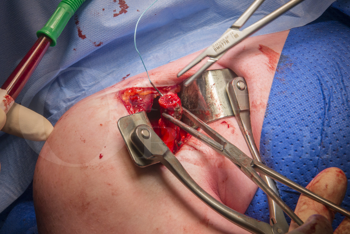
Once the decision has been made to use the technique for either primary or revision surgery then an anterior deltopectoral approach is used.Once the coracoid bone graft is harvested it is rotated through a split in subscapularis and fixed onto the prepared anterior glenoid with two partially threaded cancellous screws.This technique describes repair of the capsulolabral soft tissues to the native glenoid on the inside of the graft.
The modified technique described here uses the congruent arc Latarjet procedure, in which the curvature of the undersurface of the coracoid which matches neatly to the concavity of the glenoid socket.The cut bony surface on the medial aspect of the coracoid is created by detaching pectoralis minor with a slither of bone.This cut bony surface is then rotated to be seated onto the prepared bony surface of the anterior glenoid.This differs from the traditional or classic Latarjet technique where the entire coracoid process is harvested and then the undersurface is decorticated to be placed flat onto the glenoid surface.This technique then allowed repair of the capsular labral structures to a stump of the coracoid acromion ligament left on the lateral edge of the coracoid autograft.
In recent years arthroscopic variations of this operation have been described.These arthroscopic techniques however often ignore the repair of the soft tissues of the capsular labral complex. There is also no doubt that even for the most experienced arthroscopist this is significantly complex and challenging surgery .
All Latarjet techniques utilise the conjoint tendon attached to the tip of the coracoid to act as a sling, thus reproducing the function of the damaged anterior band of the inferior glenohumeral ligament.As the repaired shoulder rotates into abduction and external rotation the position of the conjoint tendon tightens with a tenodesis effect to further prevent anterior translation of the proximal humerus over the glenoid.
The technique I describe here, I believe is safe, reproducible and gives really very good results of stabilising a shoulder.There have been several publications highlighting potential risks and complications of the Latarjet stabilisation technique but I believe that the technique described here when used regularly and carefully is at low risk of producing such complications.
Author:Mr Mark Crowther, FRCS (Tr & Orth).
Institution: The Avon Orthopaedic centre, Southmead Hospital, Bristol, UK.
Surgical Steps
Welcome to the surgical steps quiz.
This tests your ability to recall the correct order in which the key steps of an operation should be performed.
There will be various legitimate variations in this order but the quiz doesn’t reflect this.
Your reference point for the steps quiz is just our operative technique.
It’s one starting point in helping you to learn the technique.
You simply “drag & drop” the boxes containing the descriptions of the steps into the order you think fits and submit for marking.
If you get it correct first time you will be presented your certificate at the end of the quiz.
If you need to do more work on the steps you access your marked version by clicking the“re-sit the quiz” button.
This will show the steps you ordered correctly highlighted in green and the incorrect ones in red.
The correct green steps are fixed and won’t move.
The red steps move differently after your initial attempt and swap locations with the step they are dragged and dropped onto .
Finally resubmit the quiz once again.
The quiz will let you keep going until you get all the steps in the right order.
The correct green steps are fixed and won’t move.
The red steps move differently after your initial attempt and swap locations with the step they are dragged and dropped onto .
Finally resubmit the quiz once again.
The quiz will let you keep going until you get all the steps in the right order.
COURSE PROGRESS
Surgical Steps
Time limit: 0
Quiz Summary
0 of 1 questions completed
Questions:
Information
You have already completed the quiz before. Hence you can not start it again.
Quiz is loading…
You must sign in or sign up to start the quiz.
You must first complete the following:
Results
Quiz complete. Results are being recorded.
Results
0 of 1 questions answered correctly
Your time:
Time has elapsed
Categories
Not categorized0%
1
Current
Review
Answered
Correct
Incorrect
Question 1 of 1
1. Question
The incision is marked on the front of the shoulder from just lateral to the palpable coracoid process (Co), running distally along the length of the proximal humerus.
Use a Cobb-like retractor to sweep the subscapularis muscle fibres from the anterior shoulder capsule.
Place a stay suture in the superior coracoid graft drill hole.
Palpation deep into the shoulder through the split in subscapularis will reveal the edge of the glenoid. Place a forked blade retractor medially onto the glenoid neck.
Use the osteotome to leave a hinge of bone at the base of the coracoid (Co).
Take a standard 2.5mm drill and place the first drill hole towards the resection end of the coracoid graft onto the original pectoralis minor surface.
Identification of structures attached to coracoid process after Kolbels retarctors have been inserted.
Place the forks of the subscapularis spreader into the incised subscapularis tendon and open to expose the anterior glenohumeral joint capsule
Lifting the arm opens the subcoracoid space. This allows digital palpation beneath the coracoid process to ensure that there is no soft tissue adhesions.
Use an osteotome to hatch-score the anterior glenoid bone.
Use the cutting diathermy blade to incise the capsule parallel to the glenoid whilst maintaining traction on the stay sutures pulling the soft tissues taut.
Using a cutting diathermy blade the coracoacromial ligament is released from the lateral coracoid by cutting down onto the surface of the Cobb-like retractor, which protects the underlying rotator cuff tendons.
Take a decent 5mm bite of anterior capsule labral soft tissues at the upper edge of the exposed capsule.
Seat the screws in the glenoid graft.
Holding the coracoid graft firmly against the glenoid insert the first drill into either of the pre-prepared drill holes on the surface of the coracoid bone graft. Then drill the second hole also.
.
Use the Kocher’s bone holding forceps to lift the coracoid process away from the base.
Index finger placed beneath the conjoint tendon from the lateral side to palpate the position of the musculocutaneous nerve.
Relax the Kobel retractor to retrieve the coracoid bone graft traction suture from underneath medial blade.
Identify the junction of the middle and inferior thirds of the subscapularis tendon and incise this transversely using a cutting diathermy blade being careful to only incise the tendon protecting the underlying anterior glenohumeral joint capsule.
With the coracoid bone graft tucked behind the medial blade the Kolbel retractors can be opened and the forked blade retractor can be replaced onto the stump of the coracoid process superiorly.
Insertion of self-retaining Jackson Burrows retractors after sharp dissection of superficial fascia and fat.
Use heavy mallet on curved osteotome for the coracoid osteotomy.
Bone nibblers are used to tidy up the coracoid graft.
Release pectoralis minor from medial coracoid.
The patient is set up in the semi-sitting position .
Dissection of the fatty streak between deltoid and pectoralis major muscles exposes the cephalic vein and the deltopectoral interval.
Position the anchors into the drill holes between the screws on the graft. Pull tightly on the sutures to reduce the capsulolabral soft tissues down to the edge of the native glenoid.
Place a Fukuda retractor into the glenohumeral joint.
Insert Kolbel’s retractors into deltopectoral interval.
Tuck the bone graft and conjoint tendon behind the medial blade of the Kolbel’s retractor.
Remove traction stay suture from graft drill hole and grasp the coracoid with Kocher’s bone holding forceps.
Clear subscapularis bursa
Use the same 2.5mm drill to create the second drill hole on the cut bony surface leaving a good bridge of bone between the two drill holes.
Use 3/0 monocryl running suture to repair the skin in the subcuticular layer.
Place the forked blade retractor on top of coracoid process to retract superior soft tissues.
Insert 2 suture anchors into the glenoid.
Remove the clip from the traction stay sutures on the anterior capsular labral structures, the pair of sutures are separated.
Use a second suture to take another bite into inferior aspect of the exposed capsule at the joint margin and place a clip on both of these stay sutures.
Remove Jackson Burrows retractors and assemble Kolbel’s retractors.